
Figure 2.
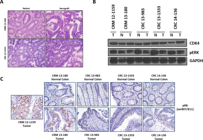
Development and characterization of a panel of CRC PDX models. (A) Histology of representative primary tumor xenografts and the patient tumor from which they were derived. (B) CDK4 and pERK expression in five individual colorectal cancer PDX models (T) and matched normal colon mucosa (N). Normal colon mucosa was not obtained from the patient for the tumor represented in lane 1 (CRM 12-1159). (C) IHC staining for pRB expression in five CRC PDX tumors and matched normal colon mucosa.