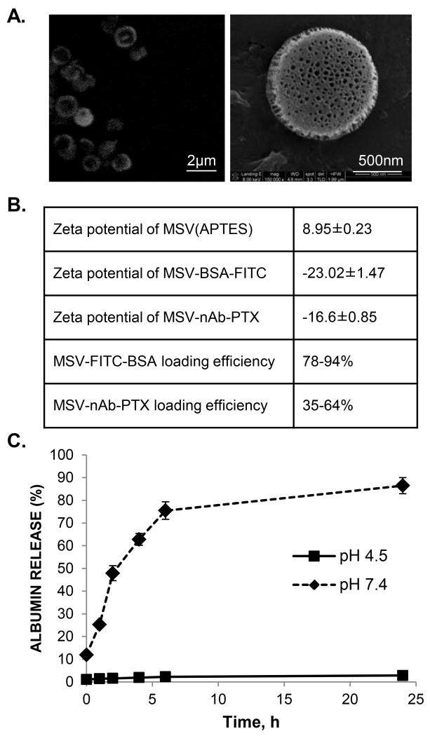
Figure 2. Loading and release of nAb-PTX in MSV.
(A) Confocal microscopic images of MSV loaded with a fluorescent probe bound to albumin (FITC-albumin) (left) and scanning electron microscopy image of MSV loaded with MSV-nAb-PTX (right); (B) Changes in zeta potential of MSV as a result of loading with FITC-albumin and nAb-PTX and loading efficiency of these molecules; (C) Release of FITC-albumin from MSV in PBS (pH 7.4) and acetic buffer (pH 4.5).

