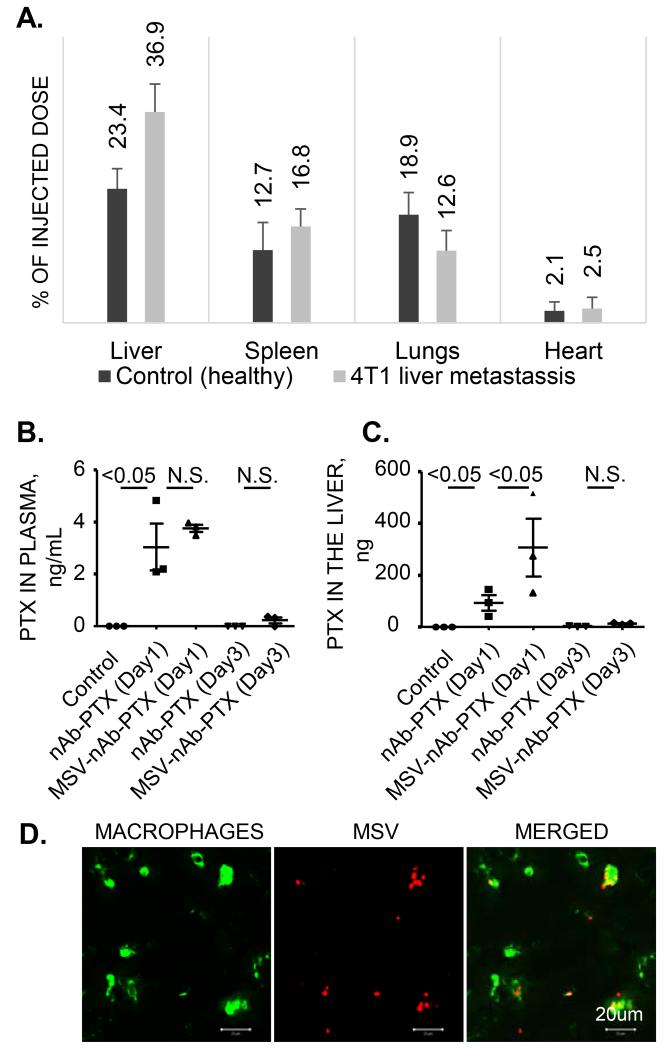
Figure 3. Biodistribution of MSV-nAb-PTX.
(A) Organ distribution of MSV injected in healthy mice and mice bearing breast cancer (4T1) lesions in the liver as determined by the elemental analysis of silicon using Inductive couples plasma atomic emission spectroscopy (ICP-AES); Levels of PTX in plasma (B) and in the liver (C), 1 and 3 days after intravenous injection of MSV-nAb-PTX and nAb-PTX into the tumor bearing mice; (D) Association of intravenously injected fluorescently labeled MSV (red) with liver macrophages (green).

