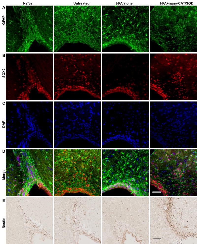
Figure 4. Immunohistochemical analysis for GFAP, SOX2, and Nestin for neuronal progenitor cells (NPCs) in the SVZ and RMS.
The NPCs could be detected with Nestin (in NPCs and transition astroglia), GFAP (in astrocytes and NPCs), and SOX2 (in non-radial neural progenitors) in IHC. The brain sections of naïve showed staining for GFAP, Nestin, and SOX2 in both SVZ and RMS. The expression of these NPCs related proteins increased after stroke as shown in the untreated sections; however, the expression of SOX2 and Nestin is reduced in RMS after the treatment with tPA alone. The treatment with tPA + nano-CAT/SOD increased the expression of GFAP, Nestin, and SOX2 than in naïve and tPA alone treated animals. Scale bar = 50 μm.

