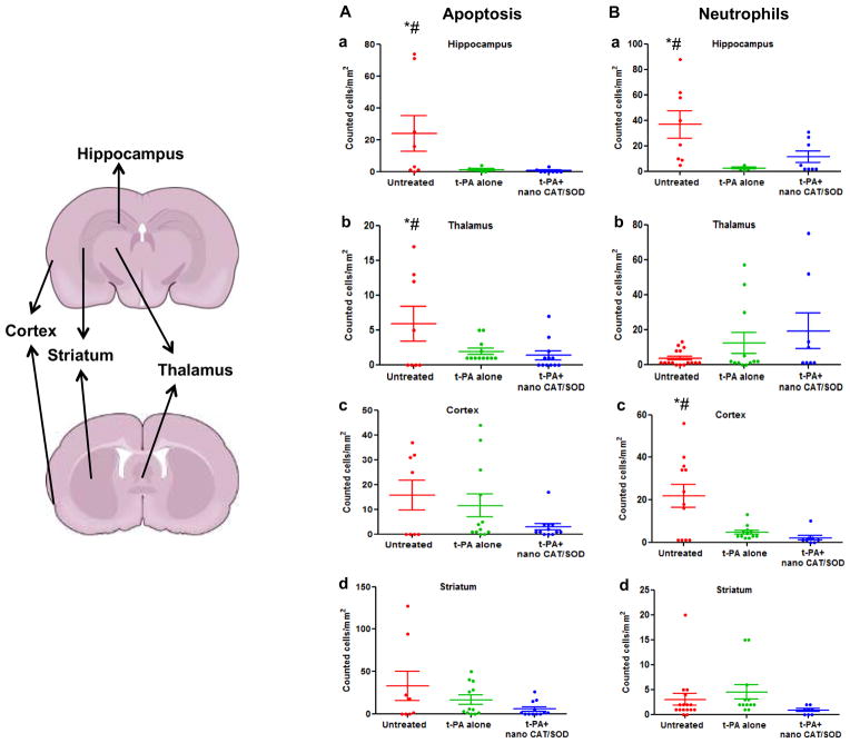
Figure 6. Quantification of apoptotic cells and infiltrating neutrophils in different regions of the brain.
Overall there is reduction in apoptotic cells in tPA+ nano-CAT/SOD treatment than in untreated and tPA alone groups. Neutrophil infiltration is also suppressed in the cortex of tPA+ nano-CAT/SOD brains vs. untreated and t-PA alone treated animal brain sections. * significant difference (p<0.05) as compared to tPA; # significant difference (p<0.05) as compared to tPA+ nano-CAT/SOD.

