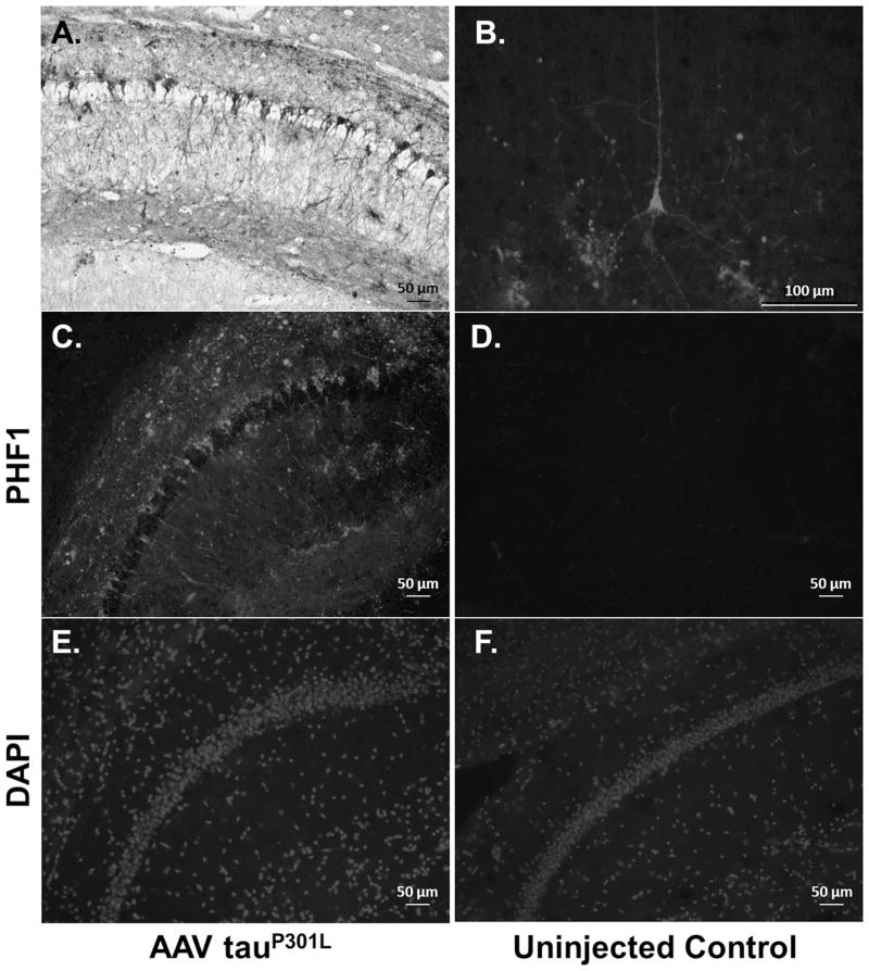
Fig. 5. Tau Expression in AAV1 TauP301L Injected Mice.
(A) AAV1 tauP301L injected brain section of a db homozygous mouse stained with PHF-1 primary antibody. Extensive tau pathology developed in the hippocampus of these mice at 6 months of age. (B) Extensive tau pathology could also be observed in the cortex. (C – F) TauP301L injected mice displayed extensive PHF1 immunoreactivity compared to uninjected controls (Top: PHF-1, Bottom: Nuclei stained with DAPI for anatomical reference). Note that no PHF1 immunoreactivity is observed in mice not injected with AAV1 tauP301L (D).

