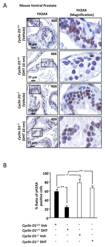
Figure 2. Cyclin D1 reduces γH2AX foci in the prostate in vivo.
(A) Immunohistochemical staining for γH2AX in the ventral prostate from cyclin D1−/− and cyclin D1+/+ mice treated with vehicle or DHT (10nm). Right panels are magnification of the inset box shown in left panels. (B) Quantitation of γH2AX positive cells from cyclin D1−/− and cyclin D1+/+ mice treated with vehicle or DHT. All data are mean ± SEM, * P<0.05 and ** P<0.01.

