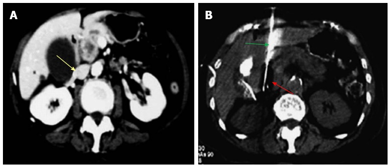
Figure 2.

Percutaneous cryoablation of pancreatic cancer under enhanced computed tomography guidance. The images were taken via the left lobe of the liver. A: The tumor at the head of the pancreas (yellow arrow) is visible with high intensity; B: The probe (green arrow) was percutaneously inserted into the tumor via the left lobe of the liver; the ice ball appears as a dark area (red arrow).
