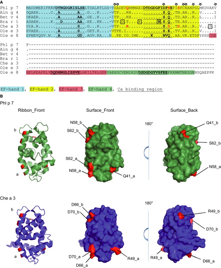
Figure 5.

(A), Protein sequence alignment of Phl p 7 with related EF‐hand allergens. Identical amino acids are indicated by dots and gaps are indicated by dashes. EF‐hand domains are coloured (EF‐hand 1: blue; EF‐hand 2: yellow; EF‐hand 3: red; EF‐hand 4: green) and calcium binding sites are bold and underscored. Amino acids that according to epitope mapping may influence binding of mAb102.1F10 are highlighted in red. Amino acids that were excluded to contribute to differences in binding are boxed or indicated by asterisks. (B) Ribbon and surface representation of Phl p 7 and Che a 3 dimers (chain a and chain b). Residues possibly involved in binding of mAb102.1F10 (see Fig. 5A, red) are highlighted on the molecular surface of Phl p 7 (green) and of Che a 3 (blue). The dimer structures are shown as ribbon and as surface representation in the same orientation (Surface_Front) and rotated 180° about the y‐axes (Surface_Back).
