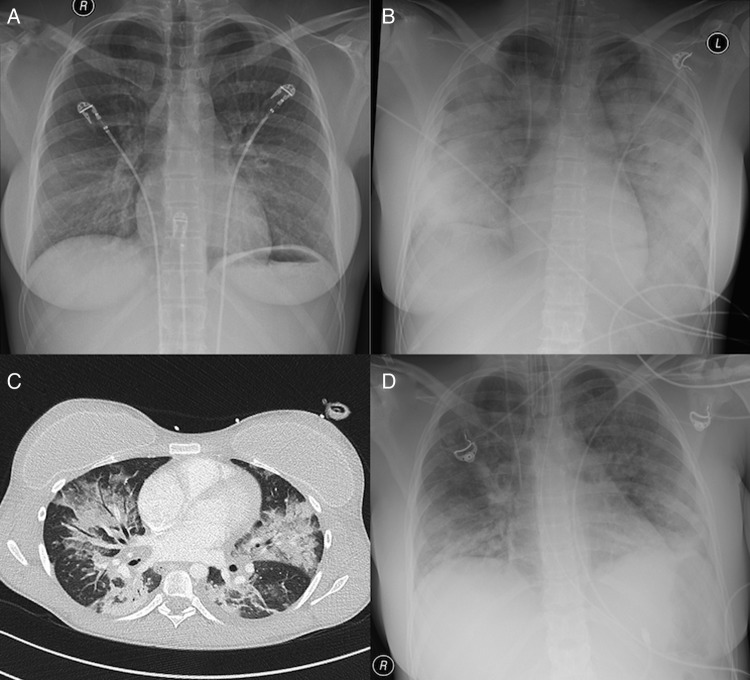
Figure 1.
Chest X-rays obtained in the emergency room on admission (A), in the intensive care unit 15 h later (B) and 1 day before extubation (D). The patchy infiltrates in (B) are consistent with a bilateral pneumonia, gradually disappearing within 14 days (D). In (C) an image obtained by CT on day 7 is shown. Bilateral extensive patchy infiltrates are shown with focal ground-glass opacities and multiple air bronchograms, again consistent with bilateral pneumonia. The patient has bilateral breast implants. There is abundant oedema fluid in the subcutaneous tissue.

