
Fig. 4.
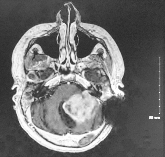
T1 with contrast MRI of patient twelve years after initial presentation, showing a heterogeneously enhancing posterior fossa mass invading the brainstem. Left sided dark signal indicates an artifact from cochlear implant.
Official websites use .gov
A
.gov website belongs to an official
government organization in the United States.
Secure .gov websites use HTTPS
A lock (
) or https:// means you've safely
connected to the .gov website. Share sensitive
information only on official, secure websites.
T1 with contrast MRI of patient twelve years after initial presentation, showing a heterogeneously enhancing posterior fossa mass invading the brainstem. Left sided dark signal indicates an artifact from cochlear implant.