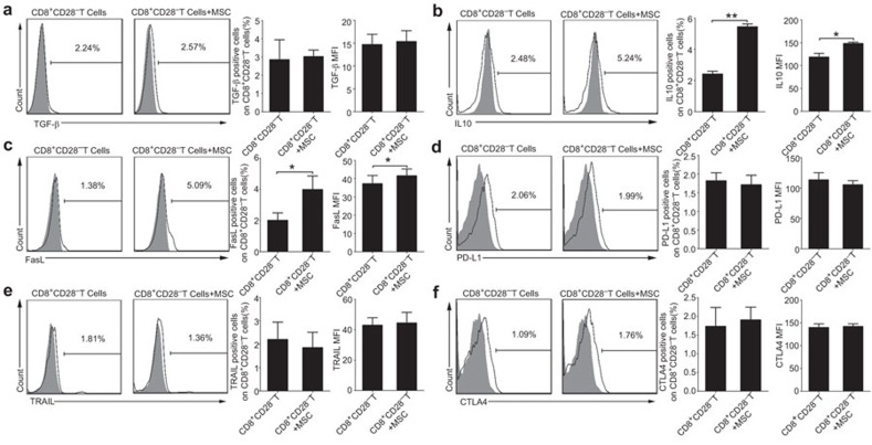
Figure 4.
MSCs affect the functions of the CD8+CD28− T cells partially by upregulating the expression of IL-10 and FasL. An analysis of the effector molecules that expressed by the CD8+CD28− T cells in the presence or absence of MSCs, including TGF-β (a), IL-10 (b), FasL (c), PD-L1 (d), TRAIL (e) and CTL-A4 (f). The frequency and mean fluorescence intensity (MFI) of IL-10 and FasL were moderately increased by MSCs (b, c). The results are representative of three independent experiments. The bar graphs indicate the means±s.d. Statistically significant differences are indicated as follows: *P<0.05 and **P<0.01. CTLA-4, cytotoxic T-lymphocyte antigen 4; MSC, mesenchymal stem cell; TRAIL, tumor necrosis factor-related apoptosis inducing ligand.

