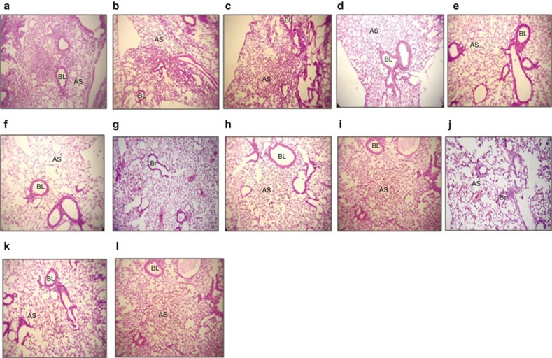
Figure 6.
Histopathology of control and immunized (rIpaB alone and rIpaB+rGroEL co-administered) mouse lung tissue. Sections of lung tissue were stained with hematoxylin and eosin and analyzed by microscopic examination. (a–c) Lung sections of control mice challenged with S. flexneri, S. boydii or S. sonnei, respectively, showing lung parenchyma with two large areas of consolidation with heavy neutrophilic cell infiltration into the lung parenchyma. (d–f) Lung sections of adjuvant control GroEL immunized mice challenged with Shigella spp. showing improved lung histology. (g–i) IpaB immunized lung section from a mouse challenged with Shigella spp. showing improved lung parenchyma with no edema in the alveolar spaces or bronchial lumina. (j–l) Lung section of a mouse co-immunized with IpaB-GroEL and challenged with Shigella spp. showing normal lung parenchyma with no inflammatory infiltrates or edema in the alveolar spaces or bronchial lumina. Images shown are at ×100 magnification. AS, alveolar space; BL, bronchial lumen.

