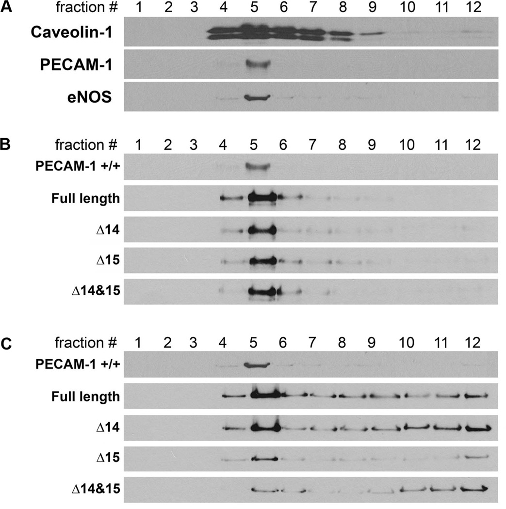
Figure 5. PECAM-1 is not sufficient for caveolae localization of eNOS.
Caveolae localization was analyzed by caveolae fractionation using sucrose gradient ultracentrifugation. Briefly, PECAM-1 +/+ (A) and PECAM-1 −/− (B, C) retinal EC expressing a specific PECAM-1 isoform and eNOS were cultured in 100 mm dishes. Confluent EC were washed and scraped off the plates with ice-cold PBS containing protease inhibitor cocktail, 3 mM Na3VO4, and 5 mM NaF and then sonicated. Protein concentration was determined using BCA protein assay kit (Pierce) and 2 ml of cell lysate (1 mg of protein) were mixed with 2 ml of 90% sucrose prepared in MES buffer (25 mM MES, 150 mM NaCl, pH 6.5) and vortexed vigorously. The sample was then added to the bottom of the centrifuge tube (Beckman, Palo Alto, CA), and 4 ml of 35% sucrose and 4 ml of 5 % sucrose in MBS (25 mM MES, 150 mM NaCl, 250 mM NaHCO3) were sequentially added. The tubes were centrifuged in a L-70 ultracentrifuge (Beckman Instrument, Palo Alto, CA) equipped with a swing rotor (SW41 Ti) at 39,000 rpm for 16 h. The gradient was separated into 1 ml fractions from the top to obtain fraction #1 (the lowest density fraction in the sucrose gradient) to 12 (the highest density fraction). A sample of each fraction was separated on SDS-PAGE and Western blotted with antibodies to caveolin-1 (BD Biosciences), anti-PECAM-1 (R&D Systems) and anti-eNOS (Santa Cruz). Retinal PECAM-1 −/− EC (B, C; express no PECAM-1 and little or no eNOS) were infected with adenovirus encoding full length, Δ14, Δ15, or Δ14&15 PECAM-1 and with eNOS, and the fractions were separated by SDS-PAGE and Western blotted with antibodies to PECAM-1 (B) or eNOS (C). Please note the caveolae localization of PECAM-1 isoforms (#5) and diffused localization of eNOS in PECAM-1 −/− EC expressing a specific PECAM-1 isoform. Although PECAM-1 caveolae localization was isoform indepepdnet, the localization of eNOS was more defuse. Full length and Δ14 isoform showed most caveolae localization. (S.Park, CM Sorenson and N. Sheibani, unpublished work)

