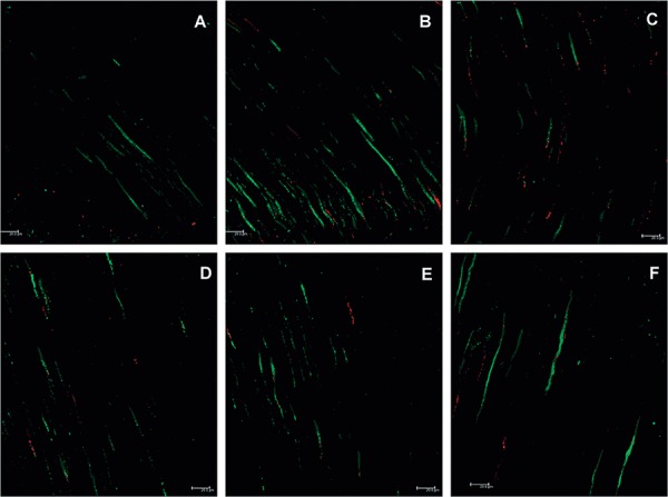
Figure 3. Confocal laser scanning microscopy (CLSM) images following group C contamination protocol. A, B, and C- Longitudinal views of the cervical third of a root canal. A greater quantity of bacteria and a predominance of live (green) bacteria can be observed; D, E, and F- Longitudinal views of the medial third of a root canal. Live bacteria can be observed. The medial third, as in all figures, is less contaminated than the cervical third.

