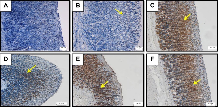
Figure 8.
HSP70 IHC.
Notes: The microscopic appearance of gastric mucosa of the rats of the normal control (A) showed insignificant HSP70 expression in the normal gastric tissue. The gastric mucosa of the rats of the ulcer control pretreated with only Tween 80 (B) showed very mild HSP70 expression in the gastric tissue (yellow arrow). However, the pretreatment with omeprazole at 20 mg/kg (C) showed increased upregulation of HSP70 expression appeared histologically as an intense brown color to the positive-stained-antigen site in the gastric tissue (yellow arrow). The pretreatment with BM at 5 mg/kg (D) showed moderate upregulation of HSP70 expression appeared histologically as an intense brown color to the positive-stained-antigen site in the gastric tissue (yellow arrow). The pretreatment with BM at 10 mg/kg (E) showed highly increased upregulation of HSP70 expression appeared histologically as an intense brown color to the positive-stained-antigen site in the gastric tissue (yellow arrow). The pretreatment with BM at 20 mg/kg (F) showed increased upregulation of HSP70 expression appeared histologically as an intense brown color to the positive-stained-antigen site in the gastric tissue (yellow arrow) (IHC: ×20).
Abbreviations: BM, β-mangostin; HSP70, heat shock protein 70; IHC, immunohistochemistry.

