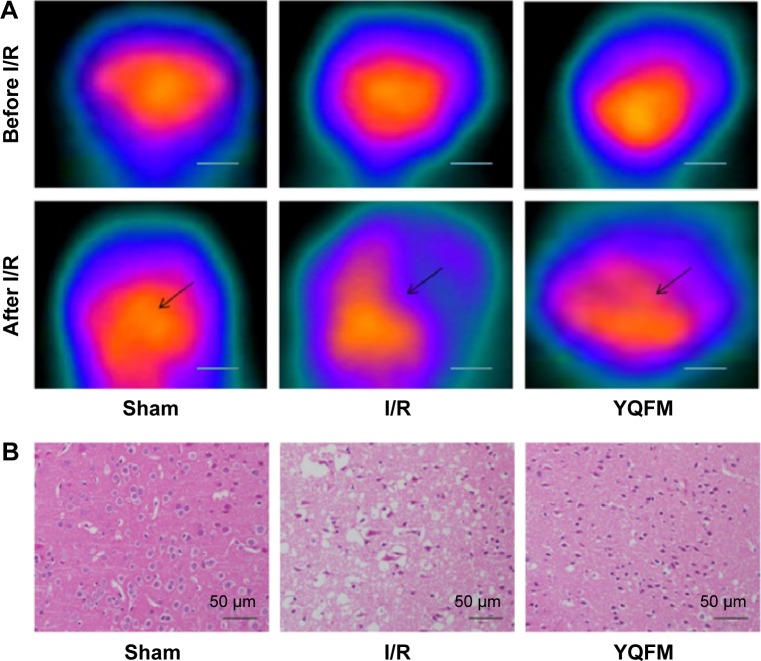
Figure 5.
Effect of YQFM on histopathological changes of brain sections in mice with cerebral I/R.
Notes: (A) (18F-FDG) PET imaging of a mouse brain before and after I/R, and representative coronal PET images of (18F-FDG) at the lesion area after surgery. The black arrows indicate the damaged region. Scale bars =200 mm. (B) Hematoxylin-and-eosin-stained slides of the brain sections of mouse in different groups were examined under a light microscope. Representative stained sections showed a gradual improvement in condensed nuclei in cortical cells in the high-dose YQFM (1.342 g/kg) treatment group. Scale bar =50 µm; n=3.
Abbreviations: 18F-FDG, 18F-fluorodeoxyglucose; I/R, ischemia–reperfusion; PET, positron emission tomography; SD, standard deviation; YQFM, YiQiFuMai powder injection.

