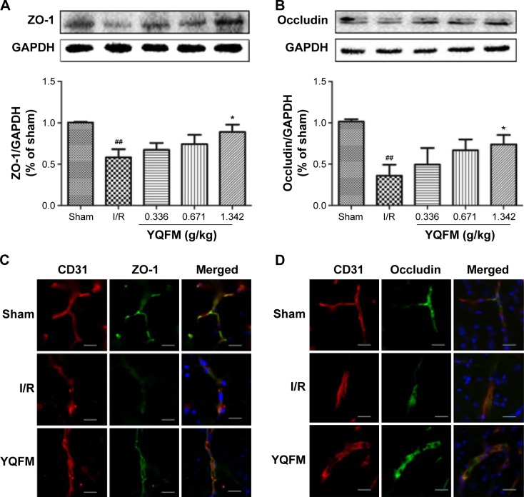
Figure 6.
Effect of YQFM on the expression of tight junction proteins in mice with cerebral I/R.
Notes: (A, B) Representative Western blots and the quantitative analysis of the ratio of ZO-1 (A) and occludin (B). (C, D) Representative immunofluorescence microscope images of ZO-1 (green) and occludin (green) localized at the periphery of endothelial cells with the marker CD31 (red). DAPI-stained nuclei are depicted in blue. Scale bars =20 µm. Distribution of ZO-1 and occludin was disrupted in the I/R group and was reduced in the high-dose YQFM (1.342 g/kg) treatment group. Data (A, B) are expressed as mean ± SD, n=3. ##P<0.01 vs sham mice; *P<0.05 vs I/R mice.
Abbreviations: GAPDH, glyceraldehyde phosphate dehydrogenase; I/R, ischemia–reperfusion; PET, positron emission tomography; SD, standard deviation; YQFM, YiQiFuMai powder injection; ZO-1, zona occludens-1.

