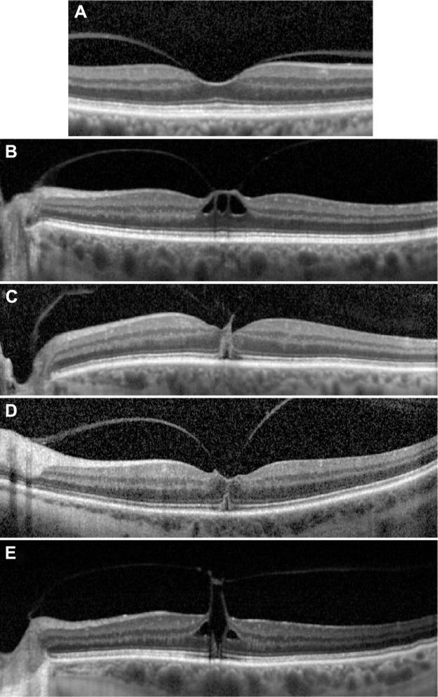
Figure 1.
SD-OCT images of Stage 1 holes.
Notes: (A) Focal VMA without traction (Grade 0). (B) VMT with inner retinal changes, (C) VMT with outer retinal changes, (D) VMT with outer retinal changes, and (E) VMT with inner and outer retinal changes.
Abbreviations: SD-OCT, spectral-domain optical coherence tomography; VMA, vitreomacular traction; VMT, vitreomacular traction.

