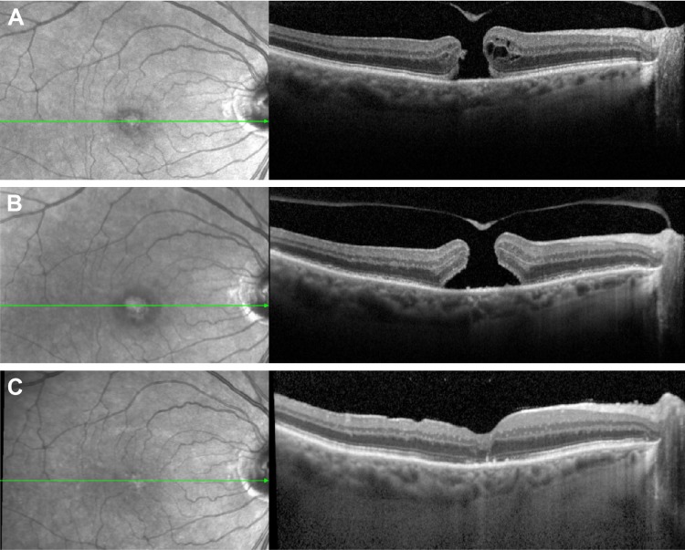
Figure 4.
SD-OCT image of a macular hole pre- and post-Ocriplasmin injection without closure.
Notes: Left, fundus photography; right, SD-OCT, green line corresponds with level of OCT image, (A) immediately pre-ocriplasmin (OCP) injection (note area of VMA was on edge of hole, not shown), (B) 1 week post OCP injection showing widened base diameter, and (C) following successful hole closure after vitrectomy and ILM peel.
Abbreviations: VMA, vitreomacular attachment; ILM, inner limiting membrane; OCP, ocriplasmin; SD-OCT, spectral-domain optical coherence tomography.

