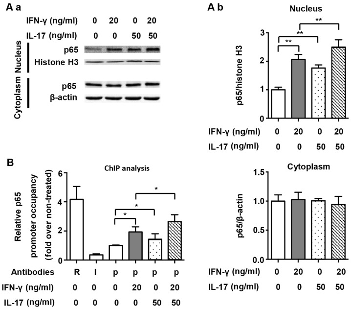
Figure 5.
Nuclear translocation and nitric oxide synthase 2 (NOS2) promoter binding of p65 by interferon-γ (IFN-γ) and/or interleukin-17 (IL-17) in RAW 264.7 cells. (A-a) Representative western blot analysis of nuclear translocation of p65 in RAW 264.7 cells treated with indicated cytokines for 1 h. β-actin was used as a cytoplasmic loading control, and histone H3 was used as a nuclear loading control. (A-b) Average quantification of nuclear and cytoplasmic p65 obtained by densitometric analysis for western blot analysis. Data are expressed as the density ratio of target protein to its non-treated level in arbitrary units. (B) Chromatin immunoprecipitation (ChIP) analysis of p65 occupancy on the proximal NF-κB element of the NOS2 promoter in RAW 264.7 cells treated with indicated cytokines for 1 h. Antibodies: R, anti-RNA Polymerase II antibody; I, IgG; p, anti-p65 antibody. Data are presented as the means ± SD from three independent experiments. *p<0.05 and **p<0.01.

