
Figure 3.
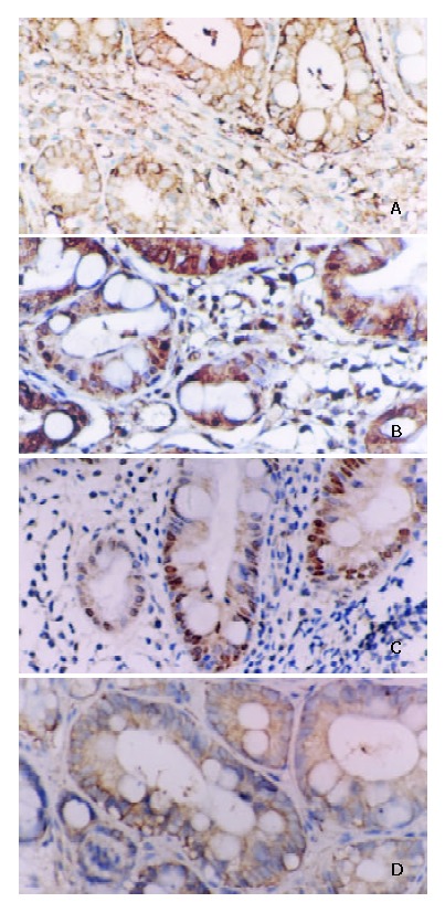
A: Positive expression of NF-κB/p65 in IM. Some inflammatory cells showed plasmatic stain. Immunohistochemical stain × 200. B: Positive expression of cyclinD1 in IM. Some inflammatory cells showed nuclear and plasmatic stains. Immunohistochemical stain × 200. C: Positive expres-sion of c-myc in IM. Some inflammatory cells showed nuclear and plasmatic stains. Immunohistochemical stain × 200. D: Positive expression of bcl-xl in IM. Some inflammatory cells showed plasmatic stain. Immunohistochemical stain × 200.