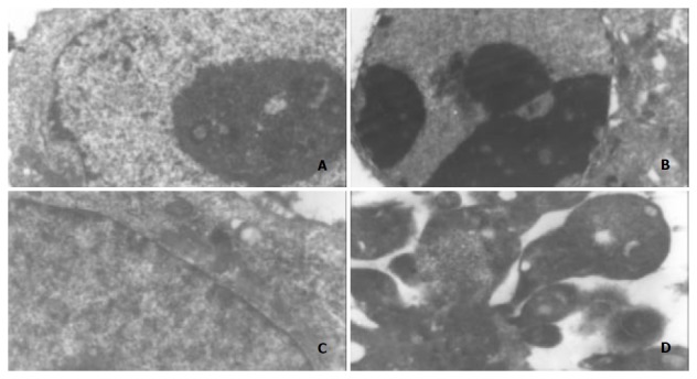
Figure 3.

Morphological changes in MGC803 and SGC7901 cells under electron microscope after treatment with ST (200 ng/ml), ×5000. A: MGC803 control cells, B: 200 ng/ml ST-induced MGC803 cells, C: SGC7901 control cells, D: 200 ng/ml ST-induced SGC7901 cells. Note: chromatin condensation and formation of apoptotic bodies.
