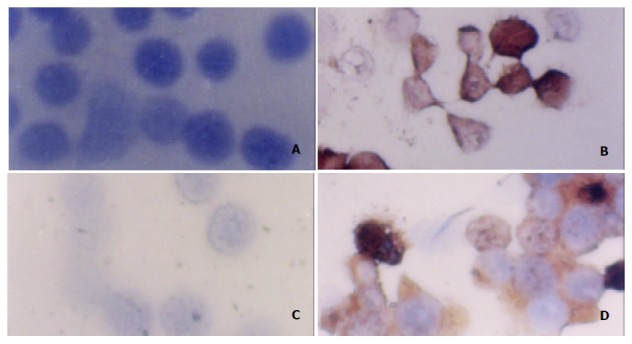
Figure 5.

Expression of P21 protein in MGC803 and SGC7901 cells after ST-treatment as determined by immunohistochemistry (SP×400). A: MGC803 control cells, B: MGC803 cells treated with 60 ng/ml ST, C: SGC7901 control cells, D: SGC7901 cells treated with 60 ng/ml ST.
