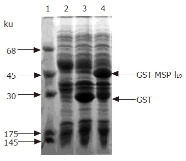
Figure 1.

Expression of fusion protein in E. coli BL21 cells. Coomassie brilliant blue-stained 12% SDS-polyacrylamide gel. lane 1, protein markers; lane 2, total protein of BL21 cells; lane 3, total protein of E. coli BL21 transformed with plasmid pGEX-5X-3 with IPTG induction; lane 4, total protein of E. coli BL21 cells harboring plasmid pGEX-MSP-119 after with IPTG induction. The arrows indicate the positions of GST and fusion protein GST-MSP-119.
