
Figure 2.
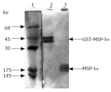
Immunoblotting analysis of MSP-119 expressed in L. lactis. Protein samples were first analyzed on 12% SDS-polyacrylamide gel and then transferred on nitrocellulose membrane followed by immunostaining with antiserum prepared by infecting mouse with P. yoelii parasites. lane 1, protein markers stained by amido black; lane 2, positive control of fusion protein GST-MSP-119 purified from E. coli cell lysate expressing the fusion protein by pGEX-MSP-119; lane 3, total protein of L. lactis cells harboring plasmid pL2-PSGT. The arrows indicate the position of fusion protein GST-MSP-119 and MSP-119.