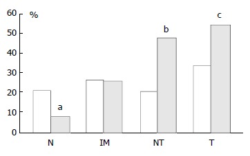
Figure 4.

Graph showing percentage of p16 positive cases as pairs of proximal (empty columns, left hand sides) and distal (black columns, right hand sides) tissue samples. N: Biopsies showing normal mucosa, IM: Biopsies showing intestinal metaplasia, NT: Non-involved mucosa from gastric cancer resection specimens, T: Tumour from gastric cancer resection specimens. There was a significant stepwise increase in expres-sion from normal mucosa→intestinal metaplasia→non-involved mucosa from cancer resections→carcinoma in the distal stomach only. aThere was a significantly lower p16 ex-pression in distal normal mucosa than in proximal normal mucosa, P = 0.0045. b and c: There was a significantly higher p16 expression in both non-involved as well as carcinoma from cancer resections from distal compared with proximal stomach, bP = 0.0048 and cP = 0.036.
