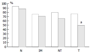
Figure 5.

Graph showing percentage of pRb positive cases as pairs of proximal (empty columns, left hand side) and distal (black columns, right hand side) tissue samples. N: Biopsies showing normal mucosa, IM: Biopsies showing intestinal metaplasia, NT: Non-involved mucosa from gastric cancer re-section specimens, T: Tumour from gastric cancer resection specimens. There was a significant stepwise decrease in ex-pression from normal mucosa→intestinal metaplasia→non-in-volved mucosa from cancer resections→carcinoma in both the distal and proximal stomach although in the latter location it was most likely due to the high expression in normal mucosa compared with the other types of tissues. aThere was a signifi-cantly lower pRb expression in distal than in proximal carcinomas, P = 0.0047.
