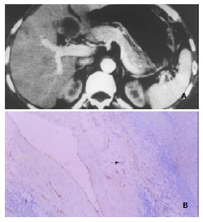
Figure 4.

Enhancement of HCC pseudocapsules and pseudocapsular MVD. A: Marked enhancement of HCC pseudocapsule (hyperdensity) in the medial segment of left hepatic lobe shown by DSCT. Note the similar enhancement pattern of the satellite or daughter lesion in the anterior seg-ment of right hepatic lobe. B: F8RA staining Rich positively stained endothelial cells within tumor pseudocapsule (black arrow) revealed by F8RA staining.
