
Figure 2.
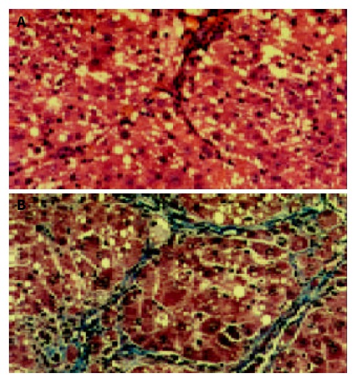
A: Liver tissue from model group showed disorderly hepatocyte cords, severe fatty degeneration, spotty or focal necrosis and infiltration of inflammatory cells. HE × 200. B: Liver tissue from model group showed collagen deposition extending from central veins or portal tracts, with thick or thin fibrotic septa and pseudolobuli formation. Masson × 200.