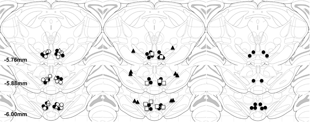
Figure 1. Intracranial injection sites.
Panels represent atlas diagrams (coronal sections −5.6, − 5.8 and −6.1 mm relative to Bregma; Paxinos and Watson, 2006) depicting injection sites from rats included in experiments in which effects of VTA injections of 1 ng/side (closed circles), 10 ng/side (open circles), or 20 ng/side (open triangles) bicuculline on CRF- and shock-induced cocaine seeking were tested (1A); rats included in experiments in which effects of VTA injections of 0.2 µg/side (closed circles) or 0.2 µg/side (open squares) 2-hydroxysaclofen on CRF- and shock-induced cocaine seeking were tested and rats with injections outside of the VTA used as anatomical controls (1B; closed triangles); and rats included in the experiments testing for effects on food-reinforced lever pressing (1C; closed circles).

