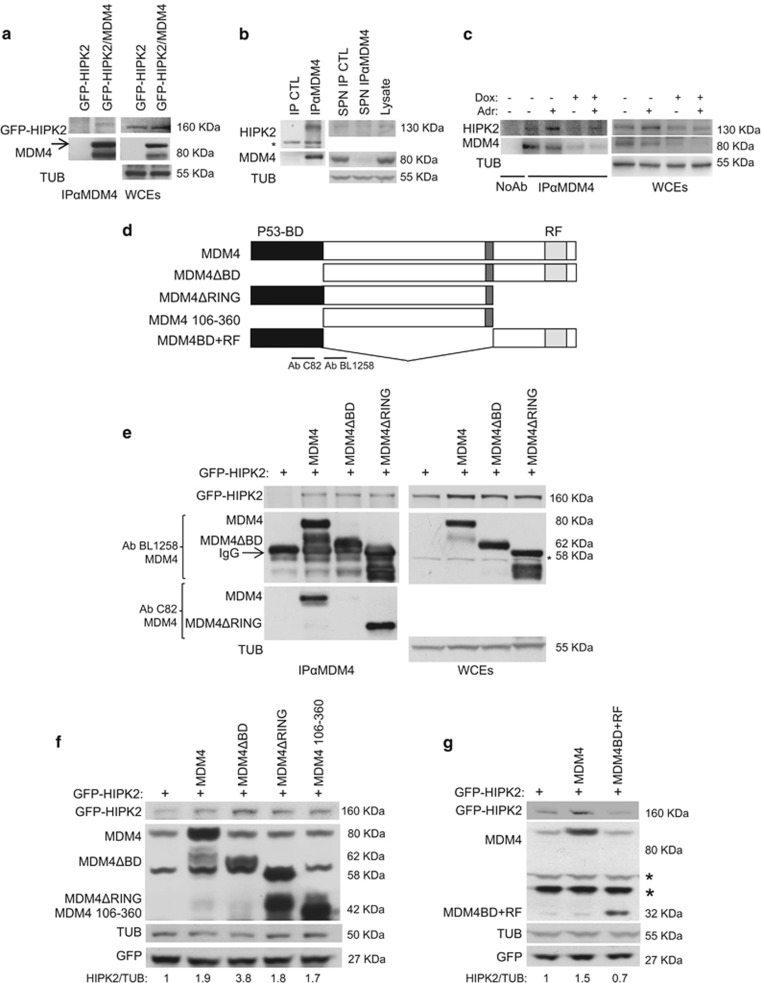
Figure 2.
MDM4-mediated stabilization of HIPK2 depends on their association. (a) Mdm4−/−p53−/− MEFs were transfected with the indicated plasmids and immunocomplexes were analysed after immunoprecipitation of 500 μg of WCE with anti-MDM4 antibody BL1258 (IPαMDM4, left panel). Right panel shows WB analysis of 1/10 of WCEs. (b) Analysis of co-immunocomplexes in MCF10A by immunoprecipitation of 2 mg of WCE with anti-MDM4 antibody BL1258 (IPαMDM4) or nonspecific IgG (IP CTL; left panel). Right panel shows WB analysis of 1/50 of WCEs (lysate). SPN IP CTL and SPN IPαMDM4 indicate supernatant of control and MDM4 immunoprecipitated samples, respectively. Asterisk (*) represents a nonspecific band. (c) Analysis of co-immunocomplexes in MCF10A-tet-shMDM4 cells treated or untreated for 24 h with doxycycline and treated with Adr for 2 h. Eight hundred micrograms of WCE were immunoprecipitated with anti-MDM4 antibody C82 (IPαMDM4). Right panel shows WB analysis of 1/20 of WCEs (lysate). (d) Scheme of MDM4 deletion mutants used in e–g. BD means p53-binding domain, RF Ring Finger domain. Underneath lines indicate the two different MDM4 antibodies used to detect deletion mutant (AbC82; AbBL1258). (e) Mdm4−/−p53−/− MEFs were transfected with the indicated plasmids and immunocomplexes analysed after immunoprecipitation of 500 μg of WCE with anti-MDM4 antibodies (IPαMDM4, left panel). The antibodies used in the WB are shown. AbC82 was used to detect MDM4ΔRING, masked by IgG in AbBL1258 blot. Right panel shows WB analysis of 1/10 of WCEs. Asterisk (*) indicates a nonspecific band. (f and g) WB of the indicated proteins in HCT116 cells transfected with the indicated plasmids. Ratio of densitometric values of HIPK2 to Tubulin is shown at the bottom of the blot. HIPK2/TUB ratio from control lane was arbitrarily set to 1.

