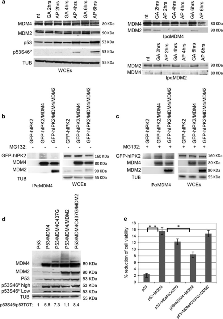
Figure 4.
MDM4 dissociates from MDM2 and promotes HIPK2/p53 functional interaction. (a) Western blot of the indicated proteins in WCEs and in co-immunoprecipitations (IP) of MCF10A cells untreated (nt) or treated with adriamycin (0.2 μM=GA) and (2 μM=AP) at the indicated time points. Immunoprecipitation were performed with anti-MDM4 antibody BL1258 (IPαMDM4, upper panel) and anti-MDM2 antibodies 2A10/Ab1 (IPαMDM2, lower panel). (b and c) Immunocomplexes of Mdm4−/−p53−/− MEFs transfected with the indicated plasmids (b) and treated with 20 μM MG132 for 6 h (c). Five hundred micrograms of WCE were immunoprecipitated with anti-MDM4 antibody BL1258 (IPαMDM4, left panels). (d) WB analysis of Mdm4−/−p53−/− MEFs transfected with indicated plasmids. Ratio of densitometric values of p53Ser46P versus p53 total levels is shown at the bottom of the blot. p53Ser46P/p53Tot ratio from p53 was arbitrarily set to 1. (e) Viability by cell titre blue colorimetric assay of cells transfected as in d. Columns represent % of reduction of cell viability in comparison with cells transfected with control vector. Mean±s.d. of two experiments in octuplicate are shown (**P<0.01; *P<0.05 Student's t-test).

