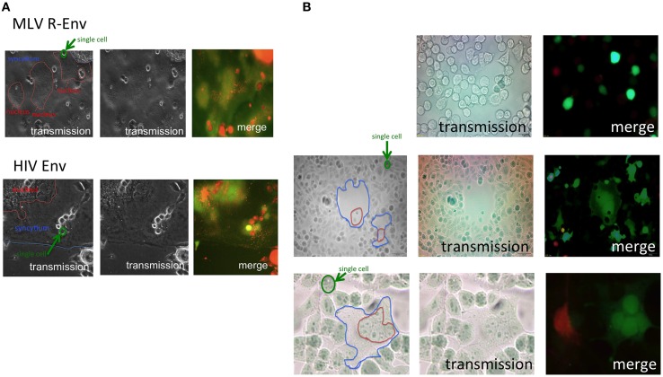
Figure 1.
Target cells are internalized into E-MLV R-Env-expressing cells. (A) Expression plasmids of the E-MLV R-Env (upper panel) or HIV-1 Env (lower panel) were transfected to HEK293T cells together with the GFP expression plasmid. TE671mCAT1 or NP2/CD4/X4 cells were stained with the cell tracker orange, and co-cultured with the transfected cells. These cells were observed under a fluorescent microscopy. Interfaces of syncytia are indicated by blue lines, and nucleic assemblies are surrounded by red lines. Single cells are surrounded by green line for scale. (B) HEK293T cells were transfected with the R+Env and GFP expression plasmids, and co-cultured with cell tracker orange-stained TE671mCAT1 cells (upper panel). HEK293T cells were transfected with the R-Env and GFP expression plasmids, and co-cultured with cell tracker orange-stained TE671 plus unstained TE671mCAT1 cells (middle and lower panels).

