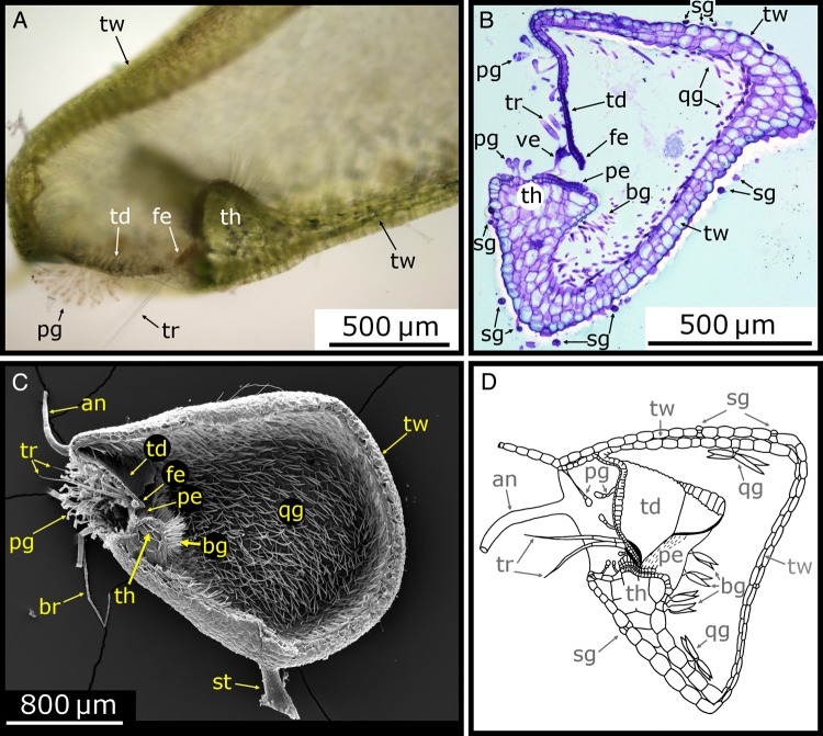
Figure 5.
Morphology of the trap body. (A) Light microscope (LM) image of a longitudinal section of a U. vulgaris trap cut open with a razor blade. The door (td) with its free edge (fe), the threshold (th), trigger hairs (tr), pyriform glands (pg) and the trap wall (tw) are visible. (B) LM image of a 10-µm-thick semi-thin longitudinal section of a U. vulgaris trap, stained with toluidine blue. The trap wall, trapdoor, threshold, spherically headed glands (sg), bifid gland (bg), quadrifid gland (qg) and the velum (ve) can be seen. (C) Scanning electron microscope image of a longitudinal section of a U. vulgaris trap (cut with a razor blade before critical point drying). Among the many structures situated at the trap entrance, especially the pavement epithelium (pe) and the bifid and quadrifid glands covering the inside of the trap are noteworthy. The stalk (st), ‘antennae’ (an) and ‘bristles’ (br) are also visible. (D) Schematic drawing of a longitudinal section of a U. gibba trap. Image modified from Lloyd (1932) (© Canadian Science Publishing or its licensors). Note that in all images, the trapdoor is arranged at an ∼90° angle to the threshold surface, which is characteristic for the U. vulgaris trap type.

