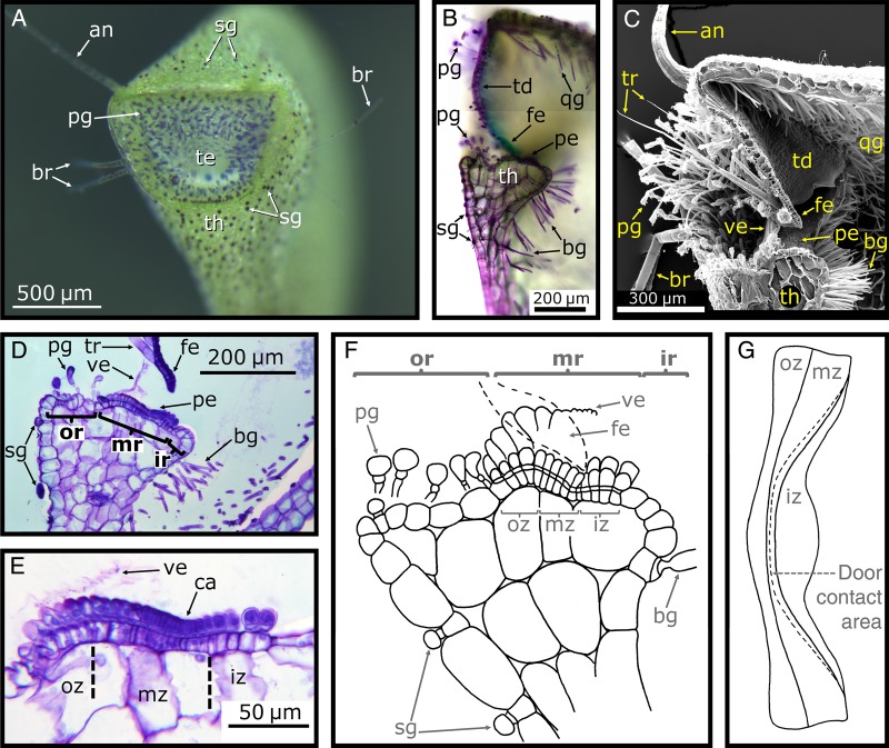
Figure 6.
Trap entrance and compartments of the threshold. (A) Inclined frontal view of a U. vulgaris trap entrance (te). After dipping the trap into toluidine blue for a few minutes, the spherically headed glands (sg) covering the outer trap surface and threshold (th) and pyriform glands (pg) at the trap entrance are very well visible. The ‘antennae’ (an) and ‘bristles’ (br) can also be seen. (B) LM image of a longitudinal section of a U. vulgaris trap entrance (cut with a razor blade), stained with toluidine blue. Note the trapdoor (td) with its free edge (fe), the pavement epithelium (pe), the bifid gland (bg) and the quadrifid gland (qg). (C) Scanning electron microscope image of a longitudinal section of a U. vulgaris trap entrance (cut with a razor blade before critical point drying); note the velum (ve). (D) LM image of a 10-µm-thick semi-thin longitudinal section of the threshold. The free door edge is also visible; note the outer (or), middle (mr) and inner (ir) region. (E) LM image of a 10-µm-thick semi-thin longitudinal section of the pavement epithelium showing the outer (oz), middle (mz) and inner (iz) zone. The door contact area, the cavity (ca), is well visible. (F) Schematic drawing of a longitudinal section of the threshold of U. gibba. The free door edge is depicted by dashed lines and is in contact with the velum, as well as with the pavement epithelium (in the cavity). (G) Schematic top view of the pavement epithelium, showing the different zones and the contact area with the door along the cavity. (F and G) Modified from Lloyd (1936a) (© Verlag Heinrich Dresden, reproduced from the Digital Library of the Royal Botanical Garden of Madrid (CSIC)).

