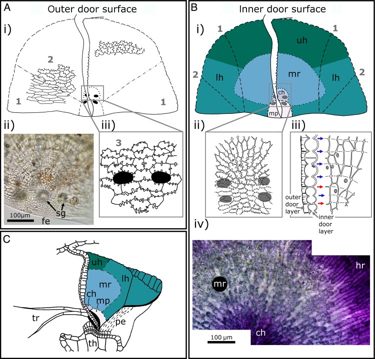
Figure 8.
Cell types on and compartmentalization of the trapdoor. (Ai) Schematic drawing of the outer door surface of a trapdoor. The course of the anticlinal cell borders is indicated. For better orientation, a schematic longitudinal section of a trapdoor is also depicted in the middle. The dashed lines highlight the transition from areas of elongated cells with anticlinal borders that run in a wavy pattern without (or with only few) reinforcing ridges (indicated by (1)) to areas of cells with zigzag anticlinal borders and pronounced reinforcing ridges (2). (Aii) LM image of the central hinge on the outer surface of a trapdoor; note the free edge (fe) and spherically headed glands (sg). The trigger hairs are out of focus. (Aiii) Schematic drawing of the central hinge (3). The insertion points of trigger hairs are highlighted by black areas. The more or less isodiametric cells of the central hinge are much smaller (compared with (1) and (2)) and possess corrugated anticlinal borders with numerous reinforcing ridges. (Bi) Schematic drawing of the inner surface of a trapdoor, and various regions (upper hinge (uh), lateral hinge (lh), middle region (mr), central hinge (ch) and middle piece (mp)) highlighted with different colours. For better orientation, a schematic longitudinal section of a trapdoor is also depicted in the middle. The insertion points of trigger hairs on the opposite outer surface of the trapdoor are marked by grey areas (surrounded by a black line in Bi)). The dashed lines (1) separate the lateral areas of the trapdoor which curve outwards and (2) depict the areas that rest on the threshold when the trapdoor is closed. (Bii and Biii) Schematic drawings of the inner cell layer. (Bii) The patterns of anticlinal borders of cells in the central hinge and in the middle piece. The cells here are small, nearly isodiametric and possess numerous pronounced reinforced ridges. Several nuclei are indicated by shaded areas. The insertion points of trigger hairs on the opposite outer surface of the trapdoor are characterized by black areas (upper image) or by grey ellipses (lower image, for better visibility). (Biii) Courses of the concentric constrictions and of the anticlinal borders of cells of the inner trapdoor layer. The left sub-image depicts a longitudinal section of the trapdoor at its middle region. The right sub-image depicts the course of the anticlinal borders. The cut cell layer on the left side visible on the right sub-image corresponds to the cells of the longitudinal section of the left sub-image. Several nuclei are indicated by shaded areas. The cells of the inner layer of the trapdoor are regularly constricted in anticlinal direction, and the constrictions correlate mostly not (blue arrows) to the transversal cellular borders (red arrows). At the areas of constrictions (indicated by short lines), cell wall reinforcements can be found. (Biv) LM image of the inner layer of a trapdoor of U. reflexa, stained with toluidine blue. The hinge region (hr), middle region and central hinge are visible. (C) Schematic drawing of the positions of the door regions (highlighted by different colours) in a ready-to-catch trap of U. gibba. The trapdoor with trigger hairs (tr) rests on the pavement epithelium (pe) of the threshold (th). Note that the lateral hinge rests on the threshold with its outer surface. (Ai, Aiii, Bi–Biii and C) Modified from Lloyd (1932). (© Canadian Science Publishing or its licensors).

