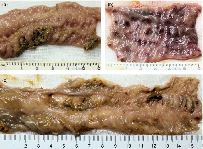
Figure 2.

Macroscopic pathology of colon. (a) Mucosal surface of normal rhesus macaque colon. (b) Mucosal surface of Shigella dysenteriae Type‐1‐infected colon at day 6 showing severe haemorrhagic gut wall damage. (c) Mucosal surface of Shigella dysenteriae‐infected colon at day 6 after dendrimer glucosamine (DG) (334 mg) treatment showing minimal damage.
