
Fig. 4.
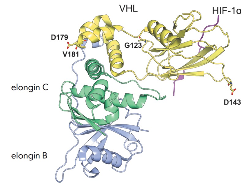
The complex of the transcription factor HIF (magenta) – von Hippel-Lindau tumor suppressor protein pVHL (yellow) – elongin C (green) – elongin B (blue). The α domain of pVHL interacts with elongin C, while the β domain binds HIF. Amino acid residues in pVHL whose motions were strongly correlated with the unstable inter-domain region are shown as sticks. The figure was prepared using PyMol based on the crystal structure 1LM8 from PDB to illustrate the results of [56]