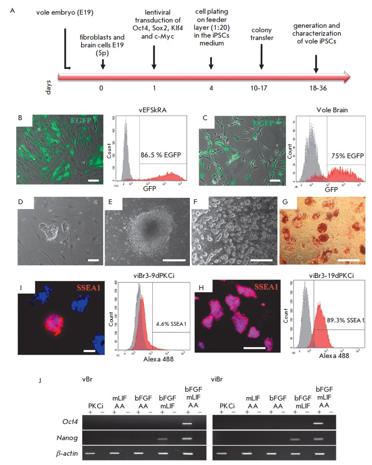
Fig. 1.

Obtaining and characterizing doxycycline-dependent iPSC-like lines of M. levis × M. arvalis hybrid cells. A – scheme of the experiment on obtaining iPSCs of the common vole of genus Microtus. B, C – assessment of the efficiency of the transduction of the embryonic skin fibroblasts (vEFSkRA cell line) and brain cells (Vole Brain) of hybrid voles with lentivirus expressing EGFP (green signal) by fluorescence microscopy and flow cytometry. The percentage of GFP-positive cells among the fibroblasts and brain cells is 86.5% and 75%, respectively. D – primary colony morphology on the 8th day of the reprogramming of cells isolated from the brain. E – colony morphology on the 13th day of reprogramming of cells isolated from the brain. F – morphology of viBr3 cell line colonies grown in the medium supplemented with mLIF + 3iR, passage 3. G – histochemical assay of endogenous AP activity in the viBr3 cell line, passage 2. H, I – data of immunofluorescence analysis and flow cytometry on the presence of SSEA1-positive cells in the viBr3 line on days 9 and 19 of culturing in the presence of PKCi, respectively. Nuclei are stained with DAPI (blue signal). J – RT-PCR analysis of Oct4 and Nanog expression in the vBr3 and viBr3 cell lines after culturing for 3 passages in the media supplemented with various components. AA – ascorbic acid. Scale bar B–D, H – 100 μm, E–G, I – 500 μm
