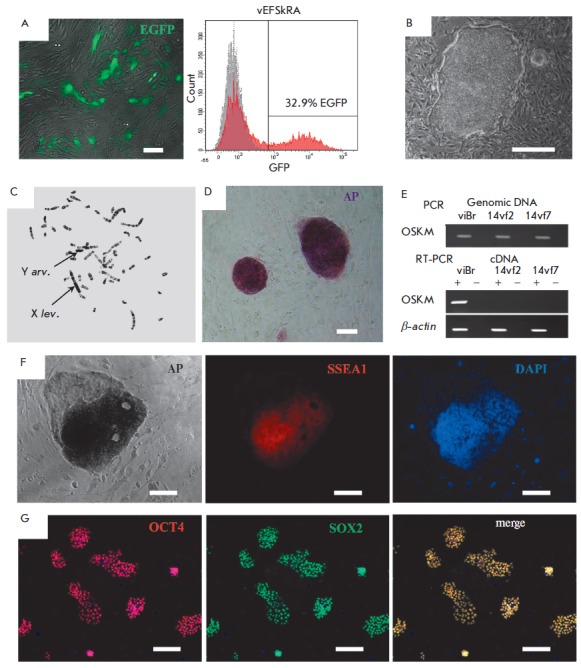
Fig. 2.

Obtaining and characterizing iPSCs of common vole M. levis × M. arvalis hybrids. A – efficiency of the transduction of vole embryonic fibroblasts (vEFSkRA) with a lentivirus expressing GFP (green signal), and assessment of the percentage of GFP-positive cells (32.9%) by fluorescence microscopy and flow cytometry. B –morphology of 14vf7 cell line colonies at passage 7. C – metaphase spread of 14vf7, passage 13. X lev. – X chromosome of M. levis, Y arv. – Y chromosome of M. arvalis. D – histochemical detection of endogenous AP activity, 14vf7 cell line, passage 6. E – RT-PCR analysis of the expression of the construct with exogenous factors of reprogramming (OSKM) in iPSC lines of common vole hybrids. F – immunofluorescence analysis of SSEA1 expression (red signal) and histochemical detection of AP activity, 14vf7 line, passage 4. Nuclei are stained with DAPI (blue signal). G – immunofluorescence analysis of pluripotency markers OCT4 (red signal) and SOX2 (green signal). Nuclei are stained with DAPI (blue signal). Scale bar A, D, F, G – 100 μm, B – 500 μm
