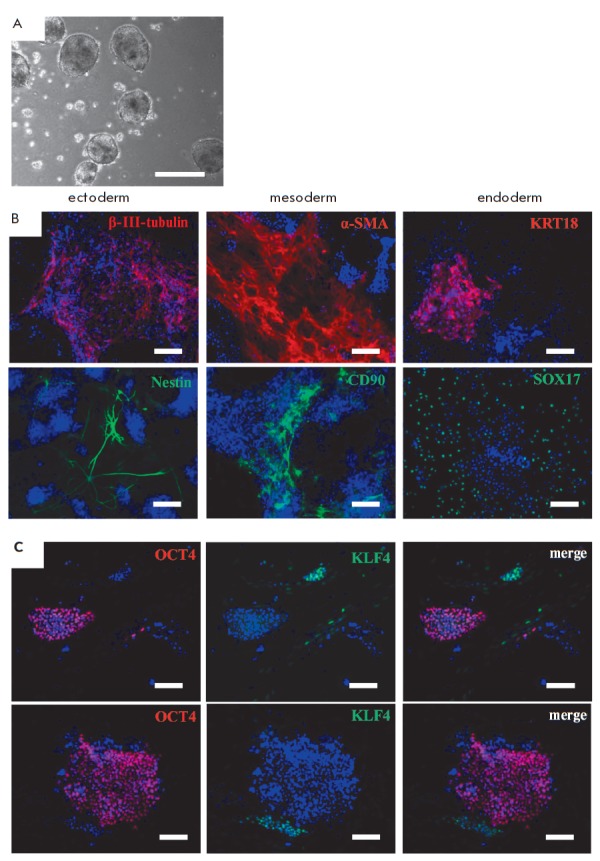
Fig. 4.

Spontaneous differentiation of common vole iPSCs. A – morphology of embryoid bodies formed from 14vf2 cells in suspension culture in 5 days. B – immunofluorescence analysis of differentiated derivatives of common vole iPSCs. Identification of ectoderm markers: β-III-tubulin (red signal), Nestin (green signal); mesoderm: α-SMA (red signal), CD90 (green signal); endoderm: KRT18 (red signal), SOX17 (green signal). C – immunofluorescent detection of the transcription factors OCT4 (red signal) and KLF4 (green signal). Nuclei are stained with DAPI (blue signal). Scale bar A – 500 μm, B, C – 100 μm
