
Figure 2.
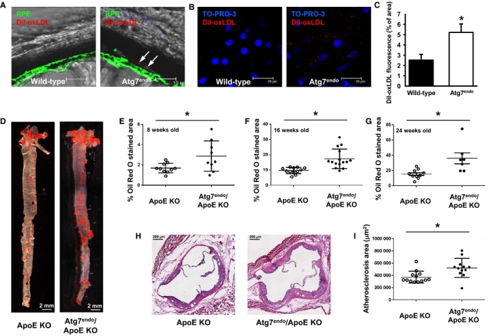
Autophagy regulates in vivo vascular lipid deposition. (A) Deposition of fluorescently labeled OxLDL in the retina of control (WT/WT; VE‐cadherin Cre) or Atg7endo mice 48 h after injection. Arrows represent retained diI‐OxLDL particles deposited sub‐RPE, at the level of the RPE basal membrane. (B) Representative whole mount images from Atg7endo mice demonstrating accumulation of fluorescent particles (presumptive vesicles) within binucleated RPE cells and in the adjacent extracellular matrix and endothelium of the choriocapillaris. (C) Quantification of labeled OxLDL in the retina of control or Atg7endo mice (n = 6 eyes per group) 48 h after infusion, *P < 0.05 by two‐tailed unpaired t‐test. (D) Representative Oil Red O stained aortas from control (WT/WT VE‐cadherin Cre/ApoE−/− abbreviated as ApoE KO) or Atg7endo/ApoE KO (fl/fl VE‐Cadherin Cre/ApoE−/−) mice. (E) Quantification of Oil Red O staining at 8 weeks (n = 10 ApoE KO and n = 9 Atg7endo/ApoE KO mice, *P < 0.05 two‐tailed unpaired t‐test with Welch's correction). (F) 16 weeks (n = 12 ApoE KO and n = 15 Atg7endoApoE KO mice, *P < 0.01 by two‐tailed unpaired t‐test with Welch's correction) and (G) 24 weeks of age (n = 10 ApoE KO and n = 7 Atg7endo/ApoE KO mice, *P < 0.03 by two‐tailed unpaired t‐test with Welch's correction). (H) Micrographs of the aortic root at 16 weeks of age. (I) Quantification of plaque area at 16 weeks between the two genotypes (n = 13 ApoE KO and n = 12 Atg7endo/ApoEKO mice, *P < 0.01 by two‐tailed unpaired t‐test with Welch's correction).