
Figure 3.
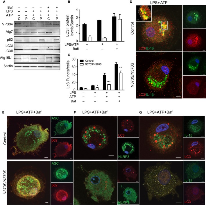
Impaired autophagy results in activation of inflammasomes in Gaucher macrophages (GMs). (A) Control (C) and GMs (P) (n = 4) were treated with lipopolysaccharide (LPS) (100 ng) or LPS+ATP (5 mm). 10 μm Baf.1A was added for 40 min after stimulation with LPS + ATP. Total lysates were run on SDS‐PAGE gels, and blots were probed with antibodies to VP34, Atg7, p62, LC3, Atg 16L1, and β‐actin. (B) LC3II protein levels normalized to β‐actin. (C) Results of a puncta assay performed in control and GMs in four independent experiments. Graph reflects the number of LC3 puncta counted per cell. Control and GMs, treated as described above, were then immunostained for (D) LC3 (red) and IL‐1β (green). (E) p62 (red) and apoptosis‐associated speck‐like proteins (green), (F) NLRP3 (green) and LC3 (red) and (G) IL‐1β (green) and LC3 (red). Images represent 20 pictures taken in 5 independent experiments (63X magnification and scale bar; 5μ).