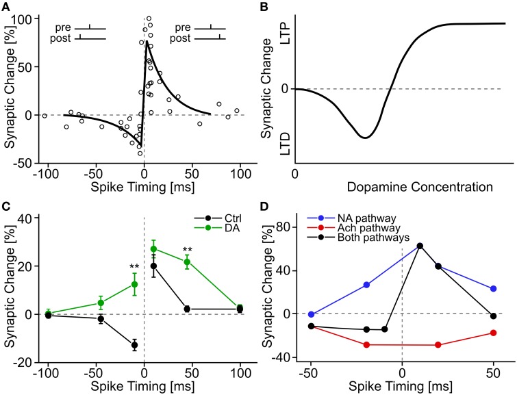
Figure 3.
Experimental evidence for STDP and neuromodulated STDP. (A) A classic example of STDP measurement. Each circle shows a measurement of synaptic change as a function of the time difference between pre- and postsynaptic spikes. The black line shows a schematic fit of an “STDP window.” Data adapted from Bi and Poo (1998), hippocampal cell culture from rat. (B) Schematic summary effect of dopamine concentration on the induction of long-term plasticity in corticostriatal synapses. Adapted from Reynolds and Wickens (2002) who summarize data from many earlier experiments. (C) Effect of extracellular dopamine (DA) on the STDP window. Adapted from Zhang et al. (2009), hippocampal cell culture from rat. (D) Effect of activation of neuromodulatory pathways on the STDP window. The noradrenaline (NA) pathway is activated through β-adrenergic receptors, the acetylcholine (Ach) pathway through M1-muscarinic receptors. Adapted from Seol et al. (2007), rat visual cortex slices.

