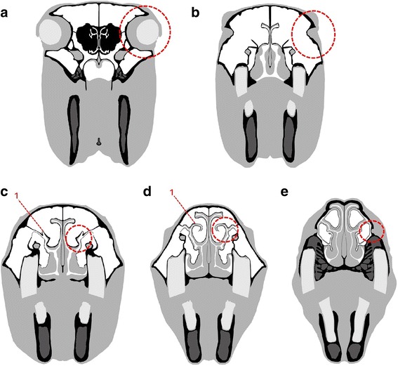
Fig. 2.

Schematic images of chosen planes. a to e Dashed circle indicates the point of orientation. a Plane 1 positioned on a plane through the centre of the ocular bulbs; b Plane 2 positioned immediately rostral to the eyes; c Plane 3 positioned on the level of the nasomaxillary aperture, a ‘hook’ (red 1) [38] protruding dorsally from the spiral lamella of the dorsal concha in all horses was used as an anatomical landmark; d Plane 4 positioned on the level of the nasomaxillary aperture, where the ‘hook’ (red 1) [38] starts to extend mediodorsally; e Plane 5 positioned on the level of the infraorbital foramen
