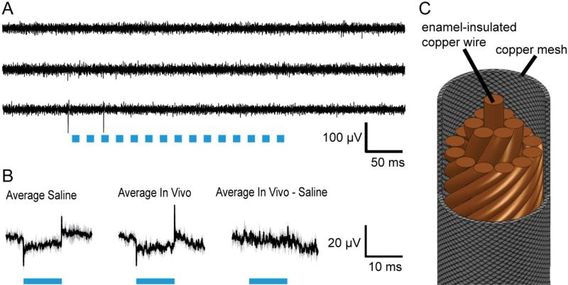
Figure 3. A custom coaxial cable design enables decoupled LED activation and recording.
A, Raw traces of three electrode recordings on a tetrode (the fourth electrode here acts as amplifier reference), of neurons in the cortex of an awake headfixed mouse (non ChR2-expressing) during 50 Hz LED operation (each pulse was for 10ms at 500mA, indicated by blue bars). B, Average, across trials and electrodes, of 33 traces obtained in each of the saline (left) and in vivo (middle) conditions, displayed as mean (solid lines) ± standard deviation between electrodes (shaded area). Right, average of the 33 traces obtained in vivo, after each trace was preprocessed by subtracting off the saline-characterized artifact recorded on the same electrode (and averaged across all trials for that electrode, e.g. 11 trials). C, Schematic of the “spatially averaged” multichannel coaxial cable utilized to deliver power to LEDs while minimizing capacitive and inductive coupling to recording electrodes.

