
Figure 3.
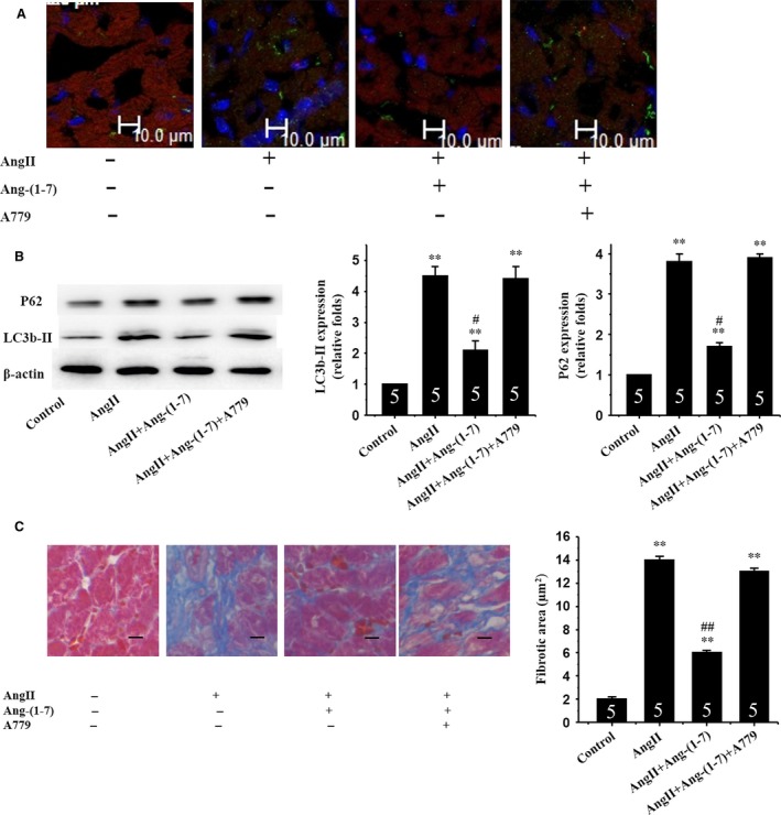
Ang‐(1‐7) inhibits AngII‐induced autophagy and myocardial fibrosis via a Mas receptor‐dependent mechanism in mice. (A) Immunofluorescent staining for expression of LC3b‐II proteins using an anti LC3b‐II antibody (green); (B) Western blot analysis for expression of LC3b‐II and P62; β‐actin in whole cell lysate used as the loading control; (C) Myocardial interstitial fibrosis. Representative Masson trichrome‐stained LV areas are shown. Blue areas indicate fibrotic staining. Fibrosis was measured in whole LV section (five sections for each mouse heart). Insets: Representative micrographs from five independent experiments. **p<0.01 vs. in the control; #p< 0.05, ##p< 0.01 vs. in the AngII‐treated mice.