
Figure 3.
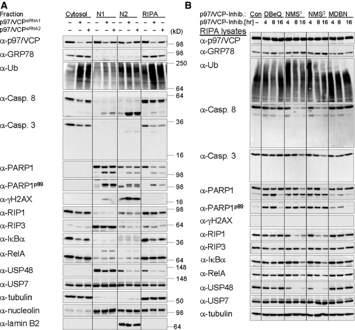
p97/VCP contributes to the maintenance of IκBα and RelA protein expression. (A) Knockdown of p97/VCP (72 hrs) induces apoptosis and reduces protein expression of IκBα. (B) Inhibition of p97/VCP with the most potent selective inhibitor NMS‐873 reduces protein expression of IκBα and RelA and induces apoptosis within hours. (A and B) Cells were harvested by subcellular fractionation or RIPA lysis, as stated, 72 hrs post RNAi of p97/VCP or at the indicated times after treatment with various p97/VCP inhibitors: DBeQ (15 μM), NMS‐873 (2.5 μM (NMS 1) or 5 μM (NMS 2)) or MDBN (15 μM). Samples were analysed by IB by use of the indicated antibodies. Tubulin (cytosol), Nucleolin (soluble nuclear fraction, N1) and Lamin B2 (insoluble nuclear fraction, N2) were used as marker proteins and/or detected for control of equal protein load. Induction of DNA damage and apoptosis was verified through IB detection of γH2AX (accumulating in N2) and/or cleavage of the nuclear protein PARP1 (detectable in N1, N2 and RIPA lysates) respectively.