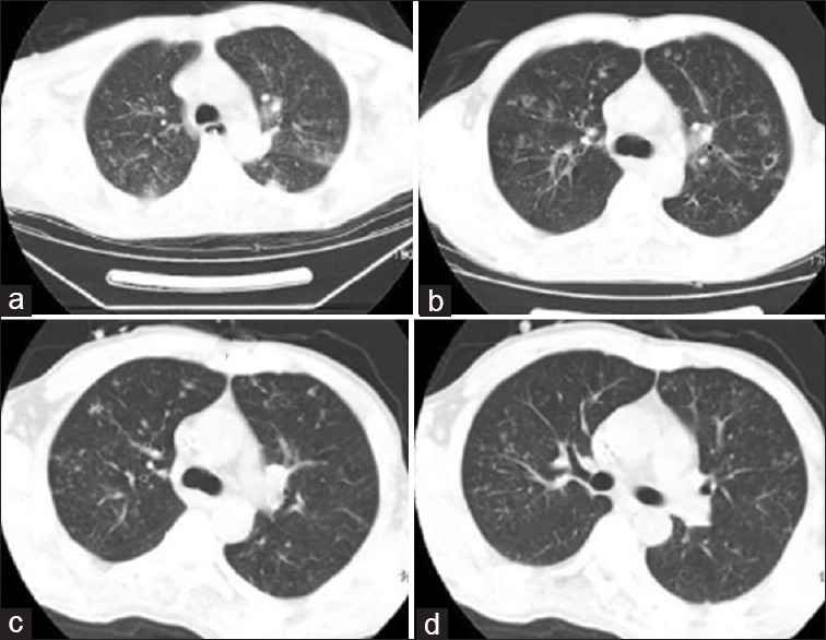
Figure 1.

Lung computed tomography findings: (a) Multiple dot- and cloud-like shadows were visible along the bronchovascular bundle. Patchy hyperdense shadows were seen at the posterior segment of the upper lobe of the left lung and the posterior segment of the lower lobes (March 7, 2008). (b) Diffuse round nodules and patchy shadows were seen in both lungs. Most nodules had small cavities with central necrosis, whereas a Halo sign was observed in a small number of nodules (March 21, 2008). (c) Diffuse round nodules and patchy shadows were seen in both lungs. The number of lesions was decreased (May 27, 2008). (d) Diffuse round nodules and patchy shadows were seen in both lungs, but became less obvious or decreased and were gradually absorbed (October 23, 2008).
